Our Technology
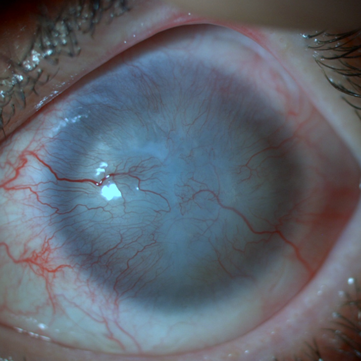
Cell therapies with the potential to treat many currently intractable diseases through uniquely powerful modes of action, have gained tremendous interest in recent years. With the development of advanced production technologies, the transfer of these technologies based on know-how to the industry, and the increasing investments in the field, cell therapy products have begun to take their place in the market. In fact, the global cell therapy market size was estimated at € 13.30 billion in 2023 and is expected to reach € 89 billion by 2033 with compound annual growth rate of 22.66% from 2024 to 2033. However, only a handful of products have been commercialised due to challenges in providing biomimetic microenvironment to the cells in vitro, non-stable production processes, and the high costs associated with their manufacturing processes. Advanced Therapy Medicinal Products (ATMPs) including cell-based tissue engineering therapies target high unmet medical needs and mainly orphan diseases such as LSCD. Limbal Stem Cell Deficiency (LSCD) is a serious ophthalmic condition that leads to corneal blindness, impacting around 375,000 patients in Europe and nearly 4 million globally. The condition results from the loss or dysfunction of limbal stem cells, which are crucial for corneal epithelium regeneration and maintenance. Without treatment, LSCD leads to vision loss, significantly affecting quality of life and the ability to participate in everyday activities.
The unique treatment of LSCD is to restore the healthy limbal stem cell pool, which has lost its activity, to the eye. Although limbal tissue transplantation is the conventional treatment option, this approach has disadvantages such as requiring a large amount (10 mm) of limbal tissue, putting the donor eye at risk for the same disease, low stem cell ratio (1-2%) and non-repeatability. To overcome these drawbacks, the methodology of in vitro cultivated limbal stem cell therapy has been developed and proposed as the most promising option. One of the most commonly used methods for in vitro expansion of limbal stem cells is culturing a 1-2 mm size of limbal biopsy on a scaffold using tissue engineering tools in the laboratory and transplanting these cells to the diseased eye. Limbustem’s founding team has improved this advanced technology using the techniques and experience they have developed (xeno-free production, using amniotic membrane and human serum, culturing with a special technique) and has already presented it to the local market as MVP. The clinical success of the current MVP is ≈ 80%. Thanks to R&D over the years, it is predicted that the stem cell content at the level of 50% is the main responsible for this clinical success. Based on the estimated positive correlation between clinical success and the stem cell content of the product, it is understood how important rising the stem cell rate will be in increasing clinical success. The team’s short-term goal for the current MVP is to increase clinical success with a fully-fledged version of the product by enhancing the stem cell ratio. In order to enhance stem cell ratio in the MVP, the team has proposed manipulating of the culture media to increase in vitro biomimicry. Tissue engineered ex vivo microenvironment must meet essential tissue-specific criteria to ensure successful integration and functional effectiveness. These criteria include high levels of biomimicry for the target tissue microenvironment; preventing immunological response; non-toxicity, cytocompatibility to promote cell growth and proliferation. Studies to date show that human amniotic membrane (hAM) is the most promising biomaterial for the expansion of limbal stem cells in vitro, as hAM does not cause immunological response, is not toxic, and moreover, naturally contains bioactive molecules such as growth factors, cytokines and extracellular matrix proteins that are vital components of native limbal niche. For instance, growth factors (e.g. EGF, NGF, FGF, IGF) regulate limbal and corneal epithelial cells haemostasis (e.g. differentiation, proliferation, migration) by mediating cellular signalling pathways in the presence of adjunct extracellular matrix components (e.g. collagens, laminin, fibronectin). hAM is a natural reservoir for these molecules. Even if hAM is the most promising biomaterial in the field, using methodology of hAM in tissue engineering (e.g. direct scaffold as tissue form), causing limited efficiency and standardization issues. Recently, idea of hAM extraction to convert it into a powder form (extract) has been proposed. In this way, drawbacks of available using methodology of hAM can be eliminated. For instance, extracted hAM’s protein content can be analysed and standardized amount of hAM content can be used as in vitro supplement. Further, powder form of hAM can be consistently supplemented into the in vitro microenvironment that provides sustainable in vitro cell expansion. For the first time in the literature, we have utilized amnion membrane extract (AME) as a supplement with standardized concentration for the explant culture of limbal stem cells. Thus, we have already shown that the addition of a biologically derived supplement that we developed allows for an increase in the ratio (≈ 70%) of stem cells in the in vitro cultured limbal epithelial cellular therapy product when compared to available MVP (≈ 50%) by preclinical analysis (unpublished data). Now, our short-term next goal is to confirm these promising outcomes with the clinical results.
Our innovation is AME supplementation into the cell media of in vitro cultured limbal epithelial cellular therapy product (Limbustem PRO AMEX).